
When ECG is the mirror of the lung Uncommon pattern in pneumothorax
Article Sidebar
Main Article Content
Abstract
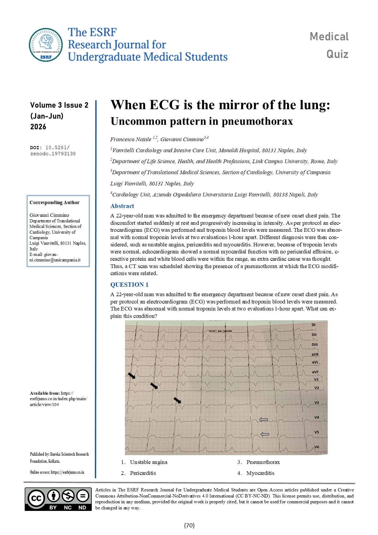
A 22-year-old man was admitted to the emergency department because of new onset chest pain. The discomfort started suddenly at rest and progressively increasing in intensity. As per protocol an electrocardiogram (ECG) was performed and troponin blood levels were measured. The ECG was abnormal with normal troponin levels at two evaluations 1-hour apart. Different diagnosis were then considered, such as unstable angina, pericarditis and myocarditis. However, because of troponin levels were normal, echocardiogram showed a normal myocardial function with no pericardial effusion, c-reactive protein and white blood cells were within the range, an extra cardiac cause was thought. Thus, a CT scan was scheduled showing the presence of a pneumothorax at which the ECG modifications were related.
Downloads
Article Details
Section

This work is licensed under a Creative Commons Attribution-NonCommercial-NoDerivatives 4.0 International License.
How to Cite


